Publié le Laisser un commentaire

Why Bone Destruction Is Accelerated with Aging

Researchers have described a relationship between the NAD+ regulator CD38, mitochondrial function, and the gradual destruction of bone in Aging Cell.

Inflammation up, bone mass down

It is well known that CD38, an enzyme that takes away NAD+, increases with age. The gradual depletion of this core metabolic molecule is, as expected, linked to multiple problems, one of which is a loss in bone-building osteoblasts [1]. Instead, CD38 contributes to the formation of osteoclasts, the cells that wear away bone [2].

Additionally, previous work has found that inflammatory conditions, including age-related inflammation (inflammaging), increases the number of monocytic myeloid-derived suppressor cells (M-MDSCs), the bone marrow cells that differentiate into osteoclasts [3]. While these researchers have found links between obesity, periodontitis, and this age-related loss of bone [4], many of the metabolic mechanisms have not been explored.

The wrong cells are accelerated with aging

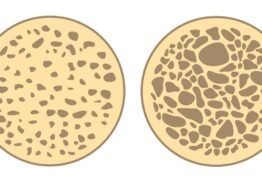
This study used six-month-old (young) and two-year-old (aged) Black 6 mice, and their tibia bones were analyzed at a cellular level. As expected, aged mice had more M-MDSCs than their younger counterparts in multiple tissues, but interestingly, there was no significant difference in the bone marrow itself, which the researchers ascribe to more migration brought about by inflammation. Not only did the aged mice have more osteoclasts, their osteoclasts were bigger and were more aggressive in wearing down bone.

This aggressiveness was accompanied by an increase in mitochondrial function. While mitochondrial dysfunction is a hallmark of aging, this did not seem to occur in these osteoclasts; instead, the osteoclasts of aged mice had strongly upregulated genes related to oxygen use and ATP production. Even the undifferentiated M-MDSCs had more mitochondrial activity in these aged animals.

This increase in activity also came with an increase in CD38. An RNA analysis found that bone marrow M-MDSCs taken from aged mice expressed eight times the CD38 as younger mice did. A more detailed gene expression analysis found that this increase in CD38 was directly correlated with genes related to accelerated bone resorption.

A small molecule, 78c, was found to be possibly effective against this increase, as it suppresses CD38. Administering 78c to the M-MDSCs derived from older mice reduced their ability to destroy bone, including the relevant mitochondrial upregulation, but it did not affect the M-MDSCs of younger mice.

This relationship between CD38, mitochondrial activity, and NAD+ is somewhat confusing and unintuitive: why would a compound that is widely known to deplete NAD+ accelerate mitochondrial function in these specific cells? It is also unclear if this activity is sex-specific, as this work was only done in male mice. Further work will have to be done to determine the role of CD38 in relation to NAD+ in these cells and to verify whether or not 78c could be safe and effective as a drug.

We would like to ask you a small favor. We are a non-profit foundation, and unlike some other organizations, we have no shareholders and no products to sell you. All our news and educational content is free for everyone to read, but it does mean that we rely on the help of people like you. Every contribution, no matter if it’s big or small, supports independent journalism and sustains our future.

Literature

[1] Kim, H. N., Ponte, F., Warren, A., Ring, R., Iyer, S., Han, L., & Almeida, M. (2021). A decrease in NAD+ contributes to the loss of osteoprogenitors and bone mass with aging. NPJ aging and mechanisms of disease, 7(1), 8.

[2] Costa, F., Toscani, D., Chillemi, A., Quarona, V., Bolzoni, M., Marchica, V., … & Giuliani, N. (2017). Expression of CD38 in myeloma bone niche: A rational basis for the use of anti-CD38 immunotherapy to inhibit osteoclast formation. Oncotarget, 8(34), 56598.

[3] Pawelec, G., Picard, E., Bueno, V., Verschoor, C. P., & Ostrand-Rosenberg, S. (2021). MDSCs, ageing and inflammageing. Cellular immunology, 362, 104297.

[4] Kwack, K. H., Zhang, L., Sohn, J., Maglaras, V., Thiyagarajan, R., & Kirkwood, K. L. (2022). Novel preosteoclast populations in obesity-associated periodontal disease. Journal of Dental Research, 101(3), 348-356.

Publié le Laisser un commentaire

40+ Experts Gather in Shanghai for TimePie Longevity Forum

From November 1st to 3rd, 2024, the 5th TimePie Longevity Forum will take place in Shanghai, China. Bringing together 40+ leading speakers from around the globe and attracting over 1,000 attendees, this event is set to be one of the most significant platforms for international anti-aging research, academic exchange, and industry collaboration in China.

Taken at the 4th TimePie Longevity Forum in 2024

A Global Dialogue on Anti-Aging Research: From Mechanisms to Translation

This gathering offers a rare opportunity to engage with a diverse array of perspectives, bringing together more than 40 leading scholars and experts from China and around the globe.

Thought leaders such as Barry Halliwell, Brian Kennedy, and Richard A. Miller will join prominent figures from China’s top universities and research institutions, including Zhao Wang from Tsinghua University, Hongbing Zhang from the Chinese Academy of Medical Sciences & Peking Union Medical College, and Yu-Xuan Lyu from The Southern University of Science and Technology. More distinguished Chinese speakers will soon be revealed.

Discussions will delve into the core mechanisms of aging, with a focus on translating the latest scientific discoveries into practical strategies for extending both healthspan and lifespan. Participants will not only gain insights into cutting-edge research but also engage in conversations about how these advancements can be applied in real-world contexts.

Connecting with China’s Expanding Longevity Market

China currently has over 260 million citizens aged 60 and above, and by 2035, this number is expected to swell to 400 million, surpassing the entire population of the United States. This dramatic demographic shift is fueling a rapidly growing demand for longevity solutions, creating a vast market ripe for innovation and development.

Amidst the diverse audience of about 1,000 participants, a significant portion—beyond the 43% academics and 39% longevity enthusiasts—comprises representatives from leading companies and investors, both domestic and international, who are deeply involved in the longevity industry.

Notably, the forum features an exceptionally international exhibitor lineup, with companies from China and around the globe spanning sectors such as CDMO, laboratories, aging diagnostics, advanced equipment, supplements, meal replacements, and traditional Chinese medicine for anti-aging.

This event serves as a crucial bridge between scientific innovation and commercial opportunity. It also provides global entrepreneurs and businesses with direct access to the Chinese market, opening doors for collaboration, investment, and expansion in one of the world’s most dynamic industries.

Immersive Experiences and Innovative Exchanges

Complementing the main discussions, the forum will feature a dynamic Biohacker Session, bringing together pioneers from China and across the globe. These trailblazers will share their bold and innovative approaches to health and longevity, sparking a vibrant exchange of ideas and experimental techniques.

Adding to the forum’s engaging atmosphere, attendees will also have the opportunity to explore a diverse exhibition area. This space will showcase the latest advancements in the anti-aging field, offering participants a chance to interact with cutting-edge products and technologies firsthand. These interactive experiences make the forum not just an intellectual gathering, but a comprehensive, hands-on exploration of the future of longevity.

The Biohacker Session at the 4th TimePie Longevity Forum

Join the 5th TimePie Longevity Forum

We believe this event is more than just a conference; it’s an opportunity to contribute to the ongoing advancements in the field of aging research.

To register and reserve your spot at this important event, please visit timepielongevityforum.com.

Join us in Shanghai to connect with leading experts and be part of a global dialogue that is helping to advance the future of longevity.

About Us

Founded by distinguished alumni from Fudan University’s Schools of Medicine, Pharmacy, and Biology, TimePie stands as a pioneering force in China’s anti-aging media landscape. Dedicated to sharing the latest breakthroughs in aging research, TimePie fosters a vibrant environment for the exchange of ideas and experiences, advancing the dialogue on aging and longevity.

Central to our mission is the annual TimePie Longevity Forum, where the world’s leading scientists, visionaries, and industry leaders gather to explore the future of aging. This event brings together experts who present groundbreaking research, alongside CEOs and biotech enthusiasts who provide valuable industry insights and share best practices.

We welcome a diverse audience, from longevity enthusiasts to biotech professionals interested in the dynamics of aging and rejuvenation. Our forum serves as a vital bridge between science and society, working towards the common goal of promoting healthier, longer lives for all.

Use code “lifespanio” for 20% off at checkout.

We would like to ask you a small favor. We are a non-profit foundation, and unlike some other organizations, we have no shareholders and no products to sell you. All our news and educational content is free for everyone to read, but it does mean that we rely on the help of people like you. Every contribution, no matter if it’s big or small, supports independent journalism and sustains our future.
Publié le Laisser un commentaire

Manuka Honey Effective Against Breast Cancer in Mice

UCLA scientists have shown that Manuka, an exclusive honey variety, can quench estrogen receptor-positive breast cancer in vitro and in vivo [1].

Honey, kill that cancer

Breast cancer remains the deadliest type of cancer in women, claiming more than 40 thousand victims a year in the US alone. Of its subtypes, estrogen receptor (ER)-positive is the most common, constituting 70-80% of all cases. Generally, ER-positive breast cancers have a better prognosis because they are often slower-growing and respond well to hormone therapy. That said, it depends on how early the cancer was detected, and the recurrence rates over several years are high.

Although modern oncology has made great strides, most anti-cancer therapies are still blunt tools that cause massive side effects and even accelerate aging [2]. The search for safer therapeutic options continues, including in the realm of natural products. Several of them, including curcumin, resveratrol, and quercetin, have shown promise against cancer, mostly in tandem with established anti-cancer therapies [3]. Honey, however, has seldom been seriously investigated.

Honey has a bad rap as “just sugar.” However, some of its varieties have been linked to a number of health benefits. Manuka honey is one of the most exclusive and expensive ones, hailing from New Zealand and some parts of Australia. It is produced by bees that pollinate the flowers of the Manuka tree (Leptospermum scoparium).

Manuka honey is known for its distinctive antibacterial properties. These are attributed mainly to the compound methylglyoxal (MGO), which is also a potent antioxidant. Other compounds abundant in Manuka honey include polyphenols and amino acids.

Recent research indicates that manuka honey can induce death by apoptosis in several cancer cell lines and helps prolong survival in some murine models of cancer [4]. Possible mechanisms of action include selective estrogen receptor modulation and inhibition of growth factor signaling pathways.

Bad for bad cells

In this new study conducted by UCLA scientists, Manuka honey and Manuka powder were first applied to cell lines of ER-positive breast cancer, triple-negative breast cancer (TNBC, a rarer but deadlier variant that lacks all three receptors common in breast cancer), and healthy mammary tissue cells.

Both honey and powder drastically decreased cellular proliferation in the ER-positive cells at high concentrations, but honey was also mildly effective in TNBC cells. In another experiment, Manuka honey was almost as effective in blocking proliferation as tamoxifen, the estrogen blocker widely used against ER-positive breast cancer. However, Manuka honey was safer for non-malignant human mammary epithelial cells (HMECs). Importantly, the two treatments showed powerful synergistic effects. Cancer cells develop resistance to tamoxifen rather quickly, making Manuka honey’s independent effectiveness important.

24 hours after treating ER-positive cancer cells with Manuka honey, more than 80% of them died by apoptosis. Here too, the effect was dose-dependent. TNBC cells also experienced apoptosis, but to a lesser degree. The researchers used a different type of honey as a control, and it did not produce any of Manuka honey’s effects.

In addition to inducing apoptosis, Manuka honey might counter the growth of cancer cells by inhibiting the growth-promoting protein mTOR. In high concentrations, Manuka honey effectively increased the levels of AMPK, a protein that negatively modulates the mTOR pathway.

Tumor growth blocked

The researchers then tested Manuka honey in a mouse model of ER-positive breast cancer. After the tumors grew to a size of 50-75 cm3, the mice started receiving orally either Manuka honey or dextrose as control, and the former blocked tumor growth almost completely.

“The findings provide hope for development of a natural, less toxic alternative to traditional chemotherapy,” said Dr. Diana Marquez-Garban, associate professor of medicine at the David Geffen School of Medicine at UCLA, and the study’s first author. “Although more research is necessary to fully understand the benefits of natural compounds in cancer therapy, this study establishes a strong foundation for further exploration in this area.”

Our findings confirm that MH has potent anticancer properties through different mechanisms. MH’s unique composition, including phenolic compounds and methylglyoxal, has been reported to have antioxidant, antiseptic, and anticancer properties. MH inhibited in vitro cell proliferation of MCF7 cells in a dose-dependent manner and induced apoptosis through PARP activation. Further, MH activated AMPK and inhibited mTOR downstream signaling as well as STAT3. Notably, orally administered MH inhibited the growth of MCF7 tumor xenografts in vivo without major side effects. These findings indicate that natural compounds such as Manuka honey, with significant antitumor activity and selectivity towards hormone receptor-positive breast cancers, may be further developed as a supplement or potential alternative to cytotoxic anticancer drugs that have more non-selective adverse effects.

We would like to ask you a small favor. We are a non-profit foundation, and unlike some other organizations, we have no shareholders and no products to sell you. All our news and educational content is free for everyone to read, but it does mean that we rely on the help of people like you. Every contribution, no matter if it’s big or small, supports independent journalism and sustains our future.

Literature

[1] Márquez-Garbán, D. C., Yanes, C. D., Llarena, G., Elashoff, D., Hamilton, N., Hardy, M., … & Pietras, R. J. (2024). Manuka Honey Inhibits Human Breast Cancer Progression in Preclinical Models. Nutrients, 16(14), 2369.

[2] Bhatia, R., Holtan, S., Jurdi, N. E., Prizment, A., & Blaes, A. (2022). Do cancer and cancer treatments accelerate aging?. Current oncology reports, 24(11), 1401-1412.

[3] Lotfi, N., Yousefi, Z., Golabi, M., Khalilian, P., Ghezelbash, B., Montazeri, M., … & Eskandari, N. (2023). The potential anti-cancer effects of quercetin on blood, prostate and lung cancers: An update. Frontiers in immunology, 14, 1077531.

[4] Ahmed, S., Sulaiman, S. A., & Othman, N. H. (2017). Oral Administration of Tualang and Manuka honeys modulates breast cancer progression in Sprague‐Dawley rats model. Evidence‐Based Complementary and Alternative Medicine, 2017(1), 5904361.