Publié le Laisser un commentaire

Engineering T Cells to Fight Brain Cancer

Researchers publishing in Nature have reported a new advance in developing chimeric antigen receptor (CAR) T cells to fight solid tumors in the brain.

A difficult endeavor

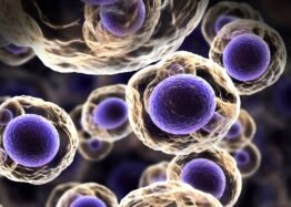
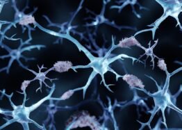
CAR T cell-based therapies are normally discussed in the context of blood cancers, as getting them to effectively attack solid tumors has proven very difficult, despite intensive research on the subject [1]. They have been used to attack glioblastomas, the most aggressive brain tumors in adults [2], even though targeting of antigens associated with this cancer can have toxic, off-target effects [3].

One safer method involves targeting of EGVRvIII, a molecule that appears on roughly 40% of these tumors [4]. However, a clinical trial utilizing this approach failed: the tumors were able to mutate and protect themselves against the treatment [5]. New approaches are being developed, and tested in trials, to better combat these tumors [6].

The researchers note the challenges involved in this sort of work. Glioblastoma tumors are very unfriendly to the immune system, suppressing its functions with increasing severity as the tumor grows [7]. For example, these tumors will secrete CD47, a natural immunosuppressant, in order to prevent macrophages from consuming them [8]. However, targeting CD47 has been found to be ineffective, as the therapy fails to penetrate the tumor, and dangerous to other tissues [9].

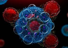
These researchers, therefore, have developed a fourth-generation therapy to target these tumors. These anti-EGFRvIII CAR T cells also release SGRP, a protein that binds to CD47, thereby directly fighting the immunosuppressive environment; however, cells also need CD47 to function properly, and the researchers were pleased to note that this alteration did not interfere with the cells’ own function.

Effective in mice

In experiments against cultured glioblastoma cells, which do not have such a protective environment, these new cells performed just as well as other CAR T cells. These cells were also found to be target-specific: cells that do not produce EGVRvIII were not harmed by these engineered cells.

In these sorts of experiments, it is relatively easy to graft human cancer cells into a mouse model and then have the CAR T cells defeat them there; however, this does not sufficiently mimic the actual tumor microenvironment, so preclinical successes can lead to clinical failures. Therefore, the researchers chose a model that avoids this problem.

The previous anti-EGFRvIII therapy was found to be effective in this scenario, extending the tumor-grafted mice’s lives and offering a one-in-five survival rate after 90 days, versus the zero they had with ineffective treatments. However, the new one performed incredibly well in comparison: after 90 days, almost none of the mice had died at all, and about two-thirds of them were completely free of tumors. The systemic toxicity associated with some forms of CAR treatment was not found in the animals treated with the new approach.

Letting other cells do their jobs

The researchers believe that some of the benefits are due to immune cell invasion of the tumors, not just of these particular T cells but of endogenous immune cells of all types. The engineered cells’ expression of SGRP within these tumors appeared to be effective. These findings were further confirmed by an analysis of cellular consumption (phagocytosis). The CAR T SGRP treatment encouraged local cells to do their jobs and consume cancer cells at a higher rate.

Spurred by their findings, the researchers also tested their SGRP approach against a mouse model of lymphoma. While they were not able to obtain the same impressive results as their glioblastoma experiment, they were able to obtain a 20% survival rate after 80 days; none of the animals treated with SGRP-less CAR T cells survived that long. While this approach did not stop lymphoma growth, it greatly slowed it down, even with just one initial treatment.

Despite sharing some of the same qualities, the researchers believe that their approach is superior to previous anti-CD47 approaches because it is expressed consistently and directly into the tumor to which the CAR T cells are attracted, a task that even locally injected antibodies have been found unable to properly do. While this is still just a mouse experiment, it may yield better clinical trial results than previous approaches.

We would like to ask you a small favor. We are a non-profit foundation, and unlike some other organizations, we have no shareholders and no products to sell you. All our news and educational content is free for everyone to read, but it does mean that we rely on the help of people like you. Every contribution, no matter if it’s big or small, supports independent journalism and sustains our future.

Literature

[1] Hou, A. J., Chen, L. C., & Chen, Y. Y. (2021). Navigating CAR-T cells through the solid-tumour microenvironment. Nature reviews Drug discovery, 20(7), 531-550.

[2] Ostrom, Q. T., Cioffi, G., Gittleman, H., Patil, N., Waite, K., Kruchko, C., & Barnholtz-Sloan, J. S. (2019). CBTRUS statistical report: primary brain and other central nervous system tumors diagnosed in the United States in 2012–2016. Neuro-oncology, 21(Supplement_5), v1-v100.

[3] Luksik, A. S., Yazigi, E., Shah, P., & Jackson, C. M. (2023). CAR T cell therapy in glioblastoma: overcoming challenges related to antigen expression. Cancers, 15(5), 1414.

[4] Felsberg, J., Hentschel, B., Kaulich, K., Gramatzki, D., Zacher, A., Malzkorn, B., … & Weller, M. (2017). Epidermal growth factor receptor variant III (EGFRvIII) positivity in EGFR-amplified glioblastomas: prognostic role and comparison between primary and recurrent tumors. Clinical Cancer Research, 23(22), 6846-6855.

[5] O’Rourke, D. M., Nasrallah, M. P., Desai, A., Melenhorst, J. J., Mansfield, K., Morrissette, J. J., … & Maus, M. V. (2017). A single dose of peripherally infused EGFRvIII-directed CAR T cells mediates antigen loss and induces adaptive resistance in patients with recurrent glioblastoma. Science translational medicine, 9(399), eaaa0984.

[6] Choi, B. D., Gerstner, E. R., Frigault, M. J., Leick, M. B., Mount, C. W., Balaj, L., … & Maus, M. V. (2024). Intraventricular CARv3-TEAM-E T cells in recurrent glioblastoma. New England Journal of Medicine, 390(14), 1290-1298.

[7] Yeo, A. T., Rawal, S., Delcuze, B., Christofides, A., Atayde, A., Strauss, L., … & Charest, A. (2022). Single-cell RNA sequencing reveals evolution of immune landscape during glioblastoma progression. Nature immunology, 23(6), 971-984.

[8] Willingham, S. B., Volkmer, J. P., Gentles, A. J., Sahoo, D., Dalerba, P., Mitra, S. S., … & Weissman, I. L. (2012). The CD47-signal regulatory protein alpha (SIRPa) interaction is a therapeutic target for human solid tumors. Proceedings of the National Academy of Sciences, 109(17), 6662-6667.

[9] Sikic, B. I., Lakhani, N., Patnaik, A., Shah, S. A., Chandana, S. R., Rasco, D., … & Padda, S. K. (2019). First-in-human, first-in-class phase I trial of the anti-CD47 antibody Hu5F9-G4 in patients with advanced cancers. Journal of Clinical Oncology, 37(12), 946-953.

Publié le Laisser un commentaire

Study of Direct Reprogramming Challenges Consensus

Scientists have shown that in a direct cellular reprogramming scenario, neurons are produced almost exclusively by a rare subtype of multipotent cells. Their findings, they claim, change our understanding of reprogramming, but not everyone agrees [1].

Direct action

In the world of cellular reprogramming, the two most well-known realms are pluripotent reprogramming, in which somatic cells are thrown back all the way into pluripotency, and partial reprogramming, in which these cells are rejuvenated without making them lose their cellular identity.

However, there is a third route called direct reprogramming, in which somatic cells are directly transformed into differentiated cells of another type [2]. A similar process happens in the body naturally in some cases, but scientists have learned to facilitate it by expressing certain factors. Transitions have been reported even between relatively distant cell types.

Both direct and pluripotent reprogramming are characterized by low efficiency, with only a fraction of the cells making the transition. There are two types of theories as to why this happens. According to one, due to multiple stochastic changes accumulated throughout cells’ lives, some of them lose the ability to transition. The other posits that from the start, only a small subset of cells has the properties necessary for transitioning.

It takes a special type of cell

In a new study published in Stem Cell Reports, researchers from the university of Toronto set out to test those theories in neonatal murine fibroblasts, which are thought to be able to transition into neurons.

The scientists obtained the cells for reprogramming from the head-and-neck region of mouse embryos. They then applied a standard protocol for direct reprogramming by virally delivering and overexpressing three neuron fate-specifying transcription factors: BRN2, ASCL1, and MYT1L, collectively known as BAM.

It is generally assumed that a large majority of cells in such a culture are murine embryonic fibroblasts (MEF) and that treating them with BAM should produce neurons, albeit with low effectiveness. What the researchers found, however, is that almost all induced neurons could be traced back to a subset of stem-like “neural crest cells”, specialized cells that originate in the developing embryo along the edges of the neural tube, the precursor to the central nervous system.

These cells can turn into a variety of cell types, including smooth muscle cells, osteoblasts, melanocytes, and fibroblasts. However, as the researchers found, they don’t disappear past the early development stage. Instead, a small number of NC cells lingers in the tissue. According to the paper, induced neurons originate almost exclusively from those NC cells.

The researchers performed several experiments to validate their findings, including the depletion of NC cells in culture, in which case, there was almost no transition into neurons.

Overturning the consensus?

This study’s results question the idea that a differentiated cell can be induced to directly transition into a distantly related cell type.

“We believed that most cases of cell reprogramming could be attributed to a rare, multi-potential stem cell that is found throughout the body and lays dormant within populations of mature cells,” said Justin Belair-Hickey, first author on the study and graduate student of U of T’s Donnelly Centre for Cellular and Biomolecular Research. “It was not fully understood why reprogramming tends to be an inefficient process. Our data explain this inefficiency by demonstrating that the neural crest stem cell is one of the few stem cells that can produce the desired reprogrammed cell type.”

“I think claims about direct reprogramming are either overstated or based on inaccurate interpretations of the data. We set out to demonstrate that the identity of a cell is much more defined and stable than the field of cellular reprogramming has proposed. At first glance, it appears that we’ve found skin cells that can be reprogrammed into neurons, but what we’ve actually found are stem cells in the skin that are derived from the brain.”

“Neural crest stem cells may have gone unnoticed by others studying cell reprogramming because, while they are widespread throughout the body, they are also rare,” said Derek van der Kooy, principal investigator on the study and professor of molecular genetics at the Donnelly Centre and U of T’s Temerty Faculty of Medicine. “As such, they may have been mistaken for mature cells of various types of tissue that could be reprogrammed into other cell types. I think what we’ve found is a unique group of stem cells that can be studied to understand the true potential of cell reprogramming.”

Relevance to the field

While their new paper focuses on direct reprogramming, the researchers mention their earlier 2019 study into pluripotent reprogramming [3], in which “a subset of MEFs exhibited an a priori propensity for reprogramming and dominance.”

However, some other researchers caution against generalizing the results. Vittorio Sebastiano, associate professor at Stanford, who is also a co-founder and SAB chair at the reprogramming-related startup Turn Biotechnologies, said to Lifespan.io: “The work is certainly interesting, but the conclusions cannot be generalized. iPSCs (induced pluripotent stem cells) can be made from many different cell types. While skin fibroblasts are poorly and loosely characterized (which may support the authors’ claims), in other reprogramming experiments, more defined and characterized cell types, such as blood cells, are used.”

“While it is important to understand the process of full reprogramming, at the end of the day, making sure you have iPSCs is what matters, and also that their pluripotency and differentiation potential are established. In summary, while understanding how the full reprogramming to pluripotency is important, these results do not really ‘cripple’ any of the findings that have been made so far.”

According to another cellular reprogramming entrepreneur, Yuri Deigin, co-founder and CEO of YouthBio Therapeutics, “these findings are specific to direct lineage reprogramming and do not impact our understanding of full pluripotent reprogramming using the Yamanaka factors.”

“Contrary to the overarching idea from the authors’ 2019 paper — that reprogramming efficiency is driven by rare ‘elite’ clones — recent work by Konrad Hochedlinger and colleagues demonstrated that transient inhibition of H3K36 methylation enables nearly 100% of somatic cells to reprogram into iPSCs,” he said. “This indicates that all cells have the potential for reprogramming when certain epigenetic barriers are overcome. While it is true that under standard conditions, only a small fraction of cells typically reach full pluripotency, this limitation is not inherent.”

We would like to ask you a small favor. We are a non-profit foundation, and unlike some other organizations, we have no shareholders and no products to sell you. All our news and educational content is free for everyone to read, but it does mean that we rely on the help of people like you. Every contribution, no matter if it’s big or small, supports independent journalism and sustains our future.

Literature

[1] Belair-Hickey, J. J., Fahmy, A., Zhang, W., Sajid, R. S., Coles, B. L., Salter, M. W., & van der Kooy, D. (2024). Neural crest precursors from the skin are the primary source of directly reprogrammed neurons. Stem Cell Reports.

[2] Wang, H., Yang, Y., Liu, J., & Qian, L. (2021). Direct cell reprogramming: approaches, mechanisms and progress. Nature Reviews Molecular Cell Biology, 22(6), 410-424.

[3] Shakiba, N., Fahmy, A., Jayakumaran, G., McGibbon, S., David, L., Trcka, D., … & Zandstra, P. W. (2019). Cell competition during reprogramming gives rise to dominant clones. Science, 364(6438), eaan0925.

Publié le Laisser un commentaire

Preventing Alzheimer’s Proteins From Accumulating

Boosting a key autophagy-related protein discourages a core component of Alzheimer’s from taking hold, according to a study published in Aging Cell.

Taking out the trash

Autophagy is the maintenance process of the cell, in which autophagosomes engulf unwanted organelles and other material and fuse together with lysosomes to be digested. As these unwanted components include such things as misfolded proteins, this is far from the first study to link autophagic deficiencies to Alzheimer’s [1, 2].

Along with the well-known amyloid beta, misfolded and modified tau is the key biomarker of Alzheimer’s disease. Tau is a necessary protein for brain function, as it provides key functions for structure and signaling [3]; however, it can also be modified in a very large number of ways, many of which lead to the death of neurons and thus cognitive decline [4]. The most well-known, and possibly most dangerous, is phosphorylation, and phosphorylated tau has been known to be core to Alzheimer’s for decades [5]. Even worse, an excess of misfolded tau can cause failures in autophagy, leading to a rapid increase in the related problems [6].

To fight back against this process and restore autophagy to distressed neurons, this research focuses on tectonin beta-propeller repeat-containing protein 1 (TECPR1), which encourages autophagosomes and lysosomes to fuse [7], accelerates the consumption of protein aggregates, including in stem cells [8], and repairs damaged lysosomes [9]. However, TECPR1 had never been previously investigated in the context of Alzheimer’s.

Tau tangles lead to impaired clearance

This study began by causing a harmful, mutated form of tau, P301S-tau, to form in HEK293 human kidney cells. They found two harmful effects: first, that P301S-tau was discouraging autophagosomes from forming in the first place and then that this form of tau was preventing autophagosomes and lysosomes from combining.

This finding was replicated in mice. Transgenic mice that expressed P301S-tau actually had more autophagosomes than wild-type mice; they were just unable to complete their jobs, being left free-floating within the cell. As the researchers expected, there was far less TECPR1 in the cells of the transgenic mice, including in hippocampal neurons, which are responsible for learning and memory; this held true whether the mice were born transgenic or transfected with a retrovirus at a young age. The levels of other autophagy-related proteins were also heavily dysregulated.

Transfecting HEK293 cells with TECPR1 appeared to do the opposite of P301S-tau. More autophagosomes were created in the TECPR1-transfected cells, and autosomal and lysosomal fusion was increased as well.

TECPR1 fights tau tangles in mice

With these positive results in hand, the researchers then turned to their mouse population. 8-month-old wild-type and P301S mice were transfected with a retrovirus that causes the overexpression of TECPR1, then studied a month later. In wild-type mice, this did nothing in terms of brain capability; there were no changes in learning ability nor behavior.

However, in the P301S group, there were a few marked changes. In the Morris water maze test, P301S mice were much slower to explore, and their memory was much worse. Transfection with TECPR1 brought these metrics much closer to those of wild-type mice. The transfection also caused benefits in object recognition; TECPR1-treated P301S mice were much better at distinguishing between new and old objects than their untreated, tau-tangled counterparts, and they had a greater ability to retain fear memories as well.

These findings were confirmed when the mice’s brains were analyzed. While TECPR1 did nothing beneficial for wild-type mice, using it to combat the mutant tau caused the neurons to stay alive and to make more connections with other neurons. Fundamental proteins that were reduced with P301S were restored with TECPR1. Overall, the researchers concluded that TECPR1 restores neuroplasticity to tau-impaired mice.

Further work found that the mechanism of action was indeed as the researchers had believed: both total tau and phosphorylated tau were reduced in the hippocampi of the P301S mice. An examination of gene expression found that TECPR1 did not affect the production of tau, only its consumption, and further work found that administering other autophagy-inhibiting compounds will prevent TECPR1 from having any positive effect.

With these results, the researchers believe that TECPR1 is a good target for treating Alzheimer’s disease. However, there are no known methods of getting more TECPR1 into the neurons of living people. To begin a clinical trial, either a gene therapy, ideally one that only targets the affected neurons, must be developed or a small molecule or nanoparticle must be found to efficiently administer TECPR1 into the affected cells or to cause them to upregulate it themselves.

We would like to ask you a small favor. We are a non-profit foundation, and unlike some other organizations, we have no shareholders and no products to sell you. All our news and educational content is free for everyone to read, but it does mean that we rely on the help of people like you. Every contribution, no matter if it’s big or small, supports independent journalism and sustains our future.

Literature

[1] Zhang, Z., Yang, X., Song, Y. Q., & Tu, J. (2021). Autophagy in Alzheimer’s disease pathogenesis: Therapeutic potential and future perspectives. Ageing research reviews, 72, 101464.

[2] Zhang, W., Xu, C., Sun, J., Shen, H. M., Wang, J., & Yang, C. (2022). Impairment of the autophagy–lysosomal pathway in Alzheimer’s diseases: pathogenic mechanisms and therapeutic potential. Acta Pharmaceutica Sinica B, 12(3), 1019-1040.

[3] Wang, J. Z., & Liu, F. (2008). Microtubule-associated protein tau in development, degeneration and protection of neurons. Progress in neurobiology, 85(2), 148-175.

[4] Li, C., & Götz, J. (2017). Tau-based therapies in neurodegeneration: opportunities and challenges. Nature Reviews Drug Discovery, 16(12), 863-883.

[5] Braak, H., & Braak, E. (1991). Neuropathological stageing of Alzheimer-related changes. Acta neuropathologica, 82(4), 239-259.

[6] Feng, Q., Luo, Y., Zhang, X. N., Yang, X. F., Hong, X. Y., Sun, D. S., … & Wang, J. Z. (2020). MAPT/Tau accumulation represses autophagy flux by disrupting IST1-regulated ESCRT-III complex formation: a vicious cycle in Alzheimer neurodegeneration. Autophagy, 16(4), 641-658.

[7] Kim, J. H., Hong, S. B., Lee, J. K., Han, S., Roh, K. H., Lee, K. E., … & Song, H. K. (2015). Insights into autophagosome maturation revealed by the structures of ATG5 with its interacting partners. Autophagy, 11(1), 75-87.

[8] Wetzel, L., Blanchard, S., Rama, S., Beier, V., Kaufmann, A., & Wollert, T. (2020). TECPR1 promotes aggrephagy by direct recruitment of LC3C autophagosomes to lysosomes. Nature communications, 11(1), 2993.

[9] Corkery, D. P., Castro‐Gonzalez, S., Knyazeva, A., Herzog, L. K., & Wu, Y. W. (2023). An ATG12‐ATG5‐TECPR1 E3‐like complex regulates unconventional LC3 lipidation at damaged lysosomes. EMBO reports, 24(9), e56841.