Publié le Laisser un commentaire

Maintaining Telomeres Extends Lifespan in Mice

A recent study has found that the overexpression of telomerase reverse transcriptase (TERT), which is a subunit of telomerase, an enzyme essential for telomere maintenance, leads to lifespan extension in mice without significant side effects [1].
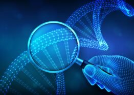
Protecting DNA

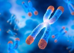
Telomere shortening is a well-known hallmark of aging. Telomeres are protective DNA sequences at the ends of the chromosomes. In most human cells, they become shorter with each division.

Telomerase and TERT have been found to be essential in maintaining telomere length [2]. Since telomeres become shorter with aging, reversing this process and extending telomere length may have the potential to extend longevity and health [3].

Creating genetically modified mice

The authors of a recent study started their research into TERT’s impact on lifespan and healthspan by creating mice that express the TERT gene. They decided to use a safer, more efficient, and more controllable approach since, as they discuss, such techniques as viruses or exogenous TERT introduction to overexpress it can “potentially lead to unintended effects or immune response,” which raises safety concerns.

The researchers genetically modified embryonic stem cells by inserting the TERT gene under the control of the human EF1α promoter. This promoter was selected to ensure stable inheritance and strong TERT expression. They referred to the genetically modified mice as TertKI.

Mating with wild-type Black 6 mice confirmed that the transgene was correctly inherited and didn’t have a negative impact on the mice’s development, growth, or survival. The researchers confirmed the transgene to be inherited for at least five generations with no negative impact on litter size.

A comparison of TertKI and wild-type animals didn’t show any significant differences in visible features, such as coat color, locomotor activities, or social behaviors, including sniffing, grooming, and play behavior.

However, the researchers noted TERT’s impact on postnatal growth and development, as the TertKI group exhibited quicker weight gain from the fifth to twenty-third day postnatal, compared to wild-type mice.

Analysis of organs revealed that organ-to-body weight ratios of the examined organs and organ cellular and tissue morphology didn’t differ between genetically modified and wild-type mice. However, analysis of organs during the autopsy revealed five cases of enlarged liver and six cases of enlarged spleen but no evidence of tumor growth.

The researchers ran tests to confirm increased TERT expression, telomerase activity, and telomere length in TertKI mice compared to wild-type mice. The results confirmed their expectations, but expression was at different levels in different organs. The authors suggested that organ-specific regulation of the EF1α promoter, TERT transcription, and/or the stability of TERT mRNA all played a role in the observed differences.

The researchers also noted that the increase in telomere length and telomerase activity in various organs was not proportional to the increase in the mRNA levels of TERT in a given organ. They suggest that this may be due to tissue-specific gene regulation.

Safety first

The researchers addressed some safety considerations regarding their research, especially since TERT gene therapy was previously debated to be either the “natural ally” or the “molecular instigator” of cancer [4]. This debate comes from the observation of telomerase activation in many human cancers.

The researchers did not observe any signs of tumors in the TertKI mice they created. Additionally, they didn’t find differences between TertKI and wild-type mice in the levels of the cancer biomarker CA72-4.

However, when the researchers exposed the mice to a mutagen to establish lung cancer, they observed more rapid cancer development in the TertKI mice compared to control animals, suggesting that the overexpression of TERT “can increase the likelihood of carcinogenesis under chronic harmful stimulation.”

Testing whether the genetic modification and TERT overexpression would cause any DNA damage or disturb fetal growth or development revealed no differences between genetically modified and wild-type mice. Blood test results either didn’t show differences or suggested that the genetically modified mice had better health.

Increased lifespan

Lifespan analysis of generations of genetically modified mice revealed an increase in the maximal lifespan of the TertKI mice by 27.48% and a 16.57% increase in median lifespan compared to WT mice.

Previous research suggested that TERT might contribute to lifespan extension through oxidative stress modulation and protection from oxidative damage, which is known to contribute to aging [5]. The researchers measured antioxidant molecules, namely glutathione (GSH) and superoxide dismutase (SOD), in mouse livers, since TERT expression was significantly increased in this organ. Both GSH and SOD were increased in the liver, suggesting improved antioxidant capacity.

However, these results might also suggest an increase in oxidative stress in TertKI mice, resulting in an increase in GSH and SOD levels. Future studies would need to address those possibilities.

Tissue repair and regenerative potential

Significant lifespan extension doesn’t seem to be the only characteristic of TertKI mice. The researchers also observed improved hair growth, faster skin wound healing with reduced infiltration of inflammatory cells, and improved collagen fiber remodeling. In vitro experiments also demonstrated that mouse TertKI skin fibroblasts had more migration ability than wild-type fibroblasts. All of these results suggest improvements in tissue repair and an increase in regenerative capacity.

An assessment of inflammatory factors during wound healing suggested a quick inflammatory response followed by a quick resolution of this inflammation. The researchers suggested that this allows for a rapid response to injury while preventing the adverse effects of an sustained inflammatory state.

The increase in the wound healing capacity of TertKI mice was also supported by the upregulation of growth factor expression and protein levels.

TERT was also found to have benefits when the researchers induced colon inflammation (colitis) in these mice. Their results indicated that their TertKI animals “display less colon deformation, functional disruption, and reduced molecular markers of injury compared to WT animals.”

Limitations

Since this study focused on the common Black 6 strain of mice, more studies are needed to test if these results are strain-specific or can be more generalizable to different strains, animal models, and environments. It is also unclear whether these findings can be applied to future human therapies in the future, especially ones that would start in older age and don’t involve TERT overexpression over the entire human lifespan.

Additionally, existng methods of overexpressing genes can be challenging to perform, time- and labor-intensive, expensive, and/or limited to mouse models. The development of easier, human therapy-compatible, and safe methods is essential.

We would like to ask you a small favor. We are a non-profit foundation, and unlike some other organizations, we have no shareholders and no products to sell you. All our news and educational content is free for everyone to read, but it does mean that we rely on the help of people like you. Every contribution, no matter if it’s big or small, supports independent journalism and sustains our future.

Literature

[1] Zhu, T. Y., Hu, P., Mi, Y. H., Zhang, J. L., Xu, A. N., Gao, M. T., Zhang, Y. Y., Shen, S. B., Yang, G. M., & Pan, Y. (2024). Telomerase reverse transcriptase gene knock-in unleashes enhanced longevity and accelerated damage repair in mice. Aging cell, e14445. Advance online publication.

[2] Bodnar, A. G., Ouellette, M., Frolkis, M., Holt, S. E., Chiu, C. P., Morin, G. B., Harley, C. B., Shay, J. W., Lichtsteiner, S., & Wright, W. E. (1998). Extension of life-span by introduction of telomerase into normal human cells. Science (New York, N.Y.), 279(5349), 349–352.

[3] Muñoz-Lorente, M. A., Cano-Martin, A. C., & Blasco, M. A. (2019). Mice with hyper-long telomeres show less metabolic aging and longer lifespans. Nature communications, 10(1), 4723.

[4] Shay J. W. (2016). Role of Telomeres and Telomerase in Aging and Cancer. Cancer discovery, 6(6), 584–593.

[5] Sahin, E., & Depinho, R. A. (2010). Linking functional decline of telomeres, mitochondria and stem cells during ageing. Nature, 464(7288), 520–528.

Publié le Laisser un commentaire

Fighting Alzheimer’s by Helping Neurons Consume Proteins

Researchers have found that kinesin family member 9 (KIF9), a protein that diminishes with aging, is instrumental in allowing cells to consume harmful proteins and fights Alzheimer’s in a mouse model.

Consuming amyloids before they become a problem

Alzheimer’s is well-known as a proteostasis disease: it is characterized by amyloid beta plaques outside the cells and tau tangles inside them [1]. These protein accumulations have been reported to occur alongside the failure of autophagy, and prior work has found that improving autophagy may be effective in preventing Alzheimer’s [2].

However, autophagy is a complicated biochemical process with many moving parts. Within neurons, the kinesin family is responsible for transporting lysosomes, core components of autophagy, along the microtubules inside the cell, and their decline is associated with brain degeneration [3]. While overexpressing kinesins have been found to improve autophagy [4], there has been little work in their connection to Alzheimer’s, and the researchers noted that KIF9 in particular has gone mostly unevaluated.

Transport is crucial

In their first experiment, the researchers examined a well-known mouse model of Alzheimer’s and compared it to wild-type mice. They found that after six months, compared to the wild-type mice, the Alzheimer’s model mice began to suffer significant reductions in KIF9 and significant increases in the proteins p62 and LCIII, which is evidence of degraded autophagy. These differences became even more stark after 12 months of life.

Next, the researchers turned to human cells. Beginning with HEK293, a commonly used cell line, the researchers used a variant, 2EB2, that produces amyloid precursor proteins. That variant, as expected, experienced diminished KIF9 and substantially decreased autophagy. This effect was found to be directional: forcing the 2EB2 cells to express more KIF9 reduced the presence of amyloid precursors and restored the autophagic components, autophagosomes, that were normally reduced in these cells.

Further research that specifically targeted individual parts of the autophagic process found that KIF9 had no special ability in directly restoring the structures themselves; rather, it was simply doing its job as a transporter, bringing these organelles to where they need to be.

Effective in mice

The researchers then used an adeno-associated virus (AAV) to determine whether an increased expression of KIF9 could ameliorate Alzheimer’s in model mice, performing behavioral tests at 5 months and brain tissue examination at 6 months.

The KIF9 AAV did nothing to the behavior of wild-type mice, as measured by an open-field test, the Barnes maze test, and the Morris water maze test. However, there were significant dfferences in all three tests when the AAV was applied to the Alzheimer’s model mice, restoring their abilities almost exactly to the levels of the wild-type mice.

Anxious mice do not want to spend time in an open space, but once they become acclimated to an area, they become more willing to explore it. Alzheimer’s model mice, at this age, do not become acclimated; the KIF9 AAV allowed this to occur significantly more.

The Barnes maze is a memory test that measures a mouse’s ability to discover and return to the correct hole. Alzheimer’s model mice, as expected, have significant impairments in memory, which the KIF9 AAV ameliorated. The Morris water maze is similar, except that it uses a hidden platform; the KIF9 AAV restored the ability of the Alzheimer’s mice to remember where it was.

However, this treatment was not perfect, as the brain examination revealed. Even after the KIF9 AAV, Alzheimer’s mice still had amyloid plaques and increases in amyloid-related proteins compared to the wild-type mice. However, the extra KIF9 did significantly reduce the amounts of these proteins and plaques.

Like many others of its kind, this is only a mouse study that uses a lab-created model, as mice do not naturally get Alzheimer’s. It is also unclear if this approach, causing neurons to express KIF9 through an AAV, could be successfully implemented in the clinic. However, it provides a crucial starting point for allowing our neurons to fight Alzheimer’s at its protein-accumulation root.

We would like to ask you a small favor. We are a non-profit foundation, and unlike some other organizations, we have no shareholders and no products to sell you. All our news and educational content is free for everyone to read, but it does mean that we rely on the help of people like you. Every contribution, no matter if it’s big or small, supports independent journalism and sustains our future.

Literature

[1] Liu, Y., Tan, Y., Zhang, Z., Yi, M., Zhu, L., & Peng, W. (2024). The interaction between ageing and Alzheimer’s disease: insights from the hallmarks of ageing. Translational Neurodegeneration, 13(1), 7.

[2] Long, Z., Ge, C., Zhao, Y., Liu, Y., Zeng, Q., Tang, Q., … & He, G. (2025). Enhanced autophagic clearance of amyloid-β via histone deacetylase 6-mediated V-ATPase assembly and lysosomal acidification protects against Alzheimer’s disease in vitro and in vivo. Neural Regeneration Research, 20(9), 2633-2644.

[3] Hayashi, K., & Sasaki, K. (2023). Number of kinesins engaged in axonal cargo transport: A novel biomarker for neurological disorders. Neuroscience Research.

[4] Liu, M., Pi, H., Xi, Y., Wang, L., Tian, L., Chen, M., … & Zhou, Z. (2021). KIF5A-dependent axonal transport deficiency disrupts autophagic flux in trimethyltin chloride-induced neurotoxicity. Autophagy, 17(4), 903-924.

Publié le Laisser un commentaire

New Study Links Epigenetic Changes to Genetic Mutations

A new paper published in Nature Aging suggests that somatic mutations cause significant remodeling of the epigenetic landscape. The findings might be relevant to future anti-aging interventions [1].

The genome and the epigenome

Genomic instability and epigenetic alterations are two of the hallmarks of aging [2]. The former occurs in somatic cells due to replication errors and stressors such as radiation and reactive oxygen species. DNA mutations can be relatively benign, but they can also impair cellular function, which might contribute to age-related disorders in various ways. The ultimate bad outcome of mutations in a single cell is, of course, cancer.

Epigenetic alterations are different. One type of them, methylation, involves a methyl group being added to or removed from a nucleotide in the DNA molecule, most often a cytosine that is followed by a guanine in the DNA sequence, with the two linked by a phosphate bond (which is why such sites are called CpG). CpG methylation is an important regulator of gene expression.

While the exact role of somatic mutations in aging is not entirely clear [3], CpG methylation is so strongly correlated with aging that it has formed the basis for epigenetic aging clocks, which have become increasingly popular over the last decade. However, what if mutations and epimutations are causally connected? A new study coming from the University of California suggests that this might be the case.

Building a mutation clock

The scientists note that at least one mechanism linking methylation and mutations has been known for a while: when a CpG site is methylated, the cytosine becomes more prone to spontaneous deamination, leading to its conversion into thymine. Since cellular DNA repair machinery does not always correct this change, CpG sites are common mutation hotspots. Conversely, if a mutation alters or eliminates a CpG site, it can prevent future methylation at that location.

Using tissue samples that had both mutation and methylation data available, the researchers identified several types of interaction between somatic mutations and DNA methylation. While they mostly used cancerous tissues, they also made an effort to validate their findings in healthy tissues.

First, the researchers confirmed that mutated CpG sites were methylated less often than non-mutated sites, which concurs with the known data. However, they also found that such mutations created atypical methylation patterns in the sections of the genome surrounding the mutation site, sometimes for tens of thousands of base pairs. This was observed in all tested tissue types.

The effect size in non-cancerous tissues, however, was substantially lower than in cancerous ones. In the latter, abnormal methylation patters were found around 15.5% of mutated sites, while in the former, the number was 8%, and the disturbances’ extent was about 1,000 base pairs from the mutation site.

Having established this correlation, the researchers wanted to see whether mutation patterns can predict biological age, just like methylation clocks do. They constructed a proprietary clock based on the profile of somatic mutations, including the counts of mutations in the vicinity of the CpG sites on which the methylation clock was based.

The methylation clock won the day, showing higher accuracy in predicting chronological age, but the mutation clock was predictive as well (Pearson correlations of r=0.83 and r=0.67, respectively). Predictions from the two clocks were also correlated across individuals. This correlation held for three previously published clocks: Horvath, PhenoAge, and Hannum.

The researchers validated their findings in a smaller number of samples from non-cancerous tissues. Here, both clocks were more predictive of chronological age (which is to be expected, since cancer introduces genomic instability and disrupts normal epigenetic patterns), but the mutation clock was still substantially behind the methylation clock. The researchers concluded that somatic mutations explain more than 50% of variation in methylation age across individuals.

What does it mean for fighting aging?

Dr. Trey Ideker of UCSD, the leading author of the study, gave us a comment:

What our paper shows is that epigenetic clocks can be largely explained by underlying DNA mutations. We think this is a pretty important finding since so much investment is currently being placed in epigenetic clocks – not only as a quantitative measurement of age, but as a means of reversing it. Our study suggests that current efforts to reverse or stabilize epigenetic changes will need to seriously contend with the underlying accumulation of DNA mutations, an area that has received comparatively less attention. On the other hand, perhaps it is worth ‘doubling down’ on treatments that slow the accumulation of DNA mutations in the first place, such as caloric restriction/dieting and certain anti-aging drugs.

The results might be especially relevant to cellular reprogramming, in which cells are being either fully de-differentiated to a pluripotent state or rejuvenated using certain reprogramming factors. Cellular reprogramming is accompanied by a considerable remodeling of the epigenetic landscape. One possibly relevant question is what if, following reprogramming, the underlying mutations cause this landscape to once again become aberrant?

“Yes, this would be one concern,” Ideker noted. “Another is that the epigenetic changes are largely not causal for aging at all, and that aging is related more directly to the mutations themselves and how they disrupt protein expression, structure and function. Essentially, what our paper has done is to open up all of these new questions.”

João Pedro de Magalhães, professor at the University of Birmingham, who was not involved in this study, said, “It’s a very interesting paper, suggesting that mutations may contribute or to some degree explain epigenetic changes, including in the context of epigenetic clocks. They show that somatic mutations with age correlate with methylation changes, which is an important new observation.”

However, he also had some reservations: “The obvious limitation of the study is that it employs data from cancer patients, including mostly from tumor samples – though some noncancerous tissues were also used. Therefore, validating these findings in normal tissues is imperative to assess the relevance of somatic mutations to epigenetic aging changes.”

One company that chose to go after the particularly hard target of fixing somatic mutations is Matter Bio. Its co-founder and CSO, Dr. Sam Sharifi, who was not involved in this study, commented:

While epigenetic clocks have attracted considerable attention as markers of biological aging, they may only reflect downstream changes triggered by a deeper, more permanent force – cumulative DNA damage. This article sheds a fascinating light on the interplay between genetic and epigenetic changes and opens the door to a purely mutation-based clock. It is still early, but once this technology matures, it could provide a more robust measure for age, given the permanent nature of DNA mutations and their steady accumulation with age.

The findings of this study are also potentially relevant to the information theory of aging promoted by Dr. David Sinclair of Harvard. It postulates that epigenetic changes are an upstream cause of aging due to loss of information on how the cell should function; therefore, aging can largely be reversed by restoring this information via cellular reprogramming or other, yet to be discovered, techniques.

“This study provides compelling evidence that epigenetic changes could not only be connected to but actually be downstream of somatic mutations,” Sharifi said. “This means that the changes in epigenetic information could be consequences of genetic information loss. Unlike methylation marks, which are relatively malleable and can be experimentally reset, DNA mutations are permanent. A big question is: are both epigenetic and genetic loss of information due to upstream processes such as DNA damage, which accumulates during aging? Critically, the article’s findings raise the important notion that targeting epigenetic states alone might not suffice to reverse aging if the underlying mutational burden is driving those epigenetic shifts in the first place.”

We would like to ask you a small favor. We are a non-profit foundation, and unlike some other organizations, we have no shareholders and no products to sell you. All our news and educational content is free for everyone to read, but it does mean that we rely on the help of people like you. Every contribution, no matter if it’s big or small, supports independent journalism and sustains our future.

Literature

[1] Koch, Z., Li, A., Evans, D. S., Cummings, S., & Ideker, T. (2025). Somatic mutation as an explanation for epigenetic aging. Nature Aging, 1-11.

[2] López-Otín, C., Blasco, M. A., Partridge, L., Serrano, M., & Kroemer, G. (2023). Hallmarks of aging: An expanding universe. Cell, 186(2), 243-278.

[3] Chatsirisupachai, K., & de Magalhães, J. P. (2024). Somatic mutations in human ageing: New insights from DNA sequencing and inherited mutations. Ageing Research Reviews, 102268.