Publié le Laisser un commentaire

A Key Protein for Tendon Health

Researchers publishing in Matrix Biology Plus have discovered that cochlin, a protein that decreases with age, is vital for the health of tendons.

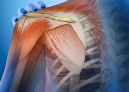
Tendons require a healthy extracellular matrix

Previous work has found that tendon tissues, which link muscle to bone, require a healthy extracellular matrix (ECM) to function; if the ECM is degraded, the tendon has problems with handling the forces put on it and in healing from injury [1]. Tenocytes, cells that maintain tendon tissue, respond to external pressures when building and removing the ECM and thus maintain tendon homeostasis [2].

These researchers have previously found that aging depletes the amount of Scleraxis-lineage tenocytes and that this depletion is directly responsible for the loss of tendon function, as demonstrated in a mouse model [3]. That work found that cochlin appears to be a crucial part of regulating tendon health.

While cochlin is found in the ECM, it is not part of structural collagen, and previous literature discussing it mostly focused on its effects in the inner ear. Its effects on tendon development have not been well documented. Therefore, this paper follows the team’s previous line of experiments, this time focusing specifically on cochlin.

Cochlin impacts how tendons mature

For their first experiment, the researchers created a strain of mice that do not produce cochlin and analyzed their tendons at 3, 6, and 9 months of age. At all three ages, the cochlin-less mice had significantly wider collagen fibrils than wild-type controls. While there were trends, there did not appear to be significant effects on how these tendons matured, except for a substantially reduced stiffness at 6 months. Peak load, however, was significantly affected: the tendons of 6-month-old cochlin-less mice could not withstand as much load as their wild-type counterparts, and this difference was only increased at 9 months.

There were also substantial alterations to tendon homeostasis, as revealed by a gene expression analysis. In line with previous research, the cochlin-less mice had signs of hearing damage, but they also had significant alterations to genes involved in protein conversion, RNA metabolism, lysosomal function, and cellular proliferation. The researchers hold that the loss of this single protein creates a broad impact to many aspects of cellular biology.

While the loss of cochlin impaired the tendons’ ability to handle stress, it did not seem to affect their ability to heal. One month after the researchers had surgically injured the flexor tendons of wild-type and cochlin-less mice at 10 to 12 weeks of age, the mice had healed in the same way. While there were trends towards decreased stiffness and decreased loading ability in the cochlin-less mice, it is unlikely that they were due to differences in how they healed. The researchers, therefore, posit that cochlin is necessary for proper tendon maturation but not healing.

Important but one of many

The researchers use the discussion section of this paper to analyze why this protein might have such wide-ranging effects. They note that it binds to collagen [4] and may have direct impacts on how its structure functions. Clearly, cochlin has impacts on maturation, but they did not attempt to analyze its effects on aging per se; as they also note, this would be particularly difficult to isolate in naturally aging animals, as a great many other things are going wrong with the ECM as well.

This work primarily notes the differences between young and middle-aged animals. Tendons, in mice, take a while to mature properly, and this paper concluded its work at almost half the animals’ natural lifespan; future work that analyzes changes with aging must therefore differentiate the positive effects of maturation versus the negative effects of age-related degeneration.

Further work that upregulates cochlin, and perhaps other ECM-related proteins that are downregulated with age, would be necessary to isolate their effects and determine whether the depletion of this protein with age is a cause or a consequence of other forms of age-related degeneration.

Collectively, these data identify Cochlin as a critical regulatory component of proper tendon structure and future work will define the therapeutic potential of conservation or restoration of Cochlin to facilitate continued tendon health through the lifespan.

We would like to ask you a small favor. We are a non-profit foundation, and unlike some other organizations, we have no shareholders and no products to sell you. All our news and educational content is free for everyone to read, but it does mean that we rely on the help of people like you. Every contribution, no matter if it’s big or small, supports independent journalism and sustains our future.

Literature

[1] Di, X., Gao, X., Peng, L., Ai, J., Jin, X., Qi, S., … & Luo, D. (2023). Cellular mechanotransduction in health and diseases: from molecular mechanism to therapeutic targets. Signal transduction and targeted therapy, 8(1), 282.

[2] Galloway, M. T., Lalley, A. L., & Shearn, J. T. (2013). The role of mechanical loading in tendon development, maintenance, injury, and repair. JBJS, 95(17), 1620-1628.

[3] Korcari, A., Nichols, A. E., Buckley, M. R., & Loiselle, A. E. (2023). Scleraxis-lineage cells are required for tendon homeostasis and their depletion induces an accelerated extracellular matrix aging phenotype. Elife, 12, e84194.

[4] Verdoodt, D., Van Camp, G., Ponsaerts, P., & Van Rompaey, V. (2021). On the pathophysiology of DFNA9: Effect of pathogenic variants in the COCH gene on inner ear functioning in human and transgenic mice. Hearing research, 401, 108162.

Publié le Laisser un commentaire

Junevity Launches to Develop Cell Reset Therapeutics

Junevity, a biotechnology company on a mission to extend lifespan and healthspan by resetting cell damage from age-related diseases, today announced $10 million in seed funding led by Goldcrest Capital and Godfrey Capital.

The Junevity RESET platform is based on exclusively licensed research by co-founder Dr. Janine Sengstack at the University of California at San Francisco. RESET uses large-scale human data and AI to identify genes – or transcription factors – that can regulate cell damage. Then, it develops siRNA therapeutics against these targets to return cells to health. Junevity will use this seed funding to enhance the RESET platform and develop its first therapeutic candidates in Type 2 diabetes, obesity and frailty.

“My research at UCSF showed the power of targeting transcription factors to restore aged human cells back to health,” said Janine Sengstack, Ph.D., co-founder and Chief Scientific Officer at Junevity. “Based on these discoveries, we are bringing forward a new class of cell reset therapeutics for diseases, with the ultimate goal of greater human longevity.”

Diseases like obesity, diabetes, frailty, neurodegeneration and many others shorten human lifespan and are associated with complex cell damage at the transcriptional level. RESET uses billions of data points from human diseases and AI to rank and evaluate potential targets. Together, the platform outputs the Cell RESET Atlas, a collection of promising transcription factor targets by cell type and by disease for therapeutic targeting. Junevity then develops novel silencing RNA (siRNA) therapeutics to restore cellular transcription back to a healthy state.

Junevity’s preclinical data demonstrates the power of the RESET platform. In Type 2 diabetes, Junevity’s first siRNA therapeutic candidate improved glucose control and insulin sensitivity in diabetic mice without causing weight gain or other side effects associated with insulin sensitizers. In obesity, Junevity’s second siRNA candidate improved adipose tissue metabolism and reduced food intake, leading to 30% weight loss versus controls. Importantly, this weight loss was driven by fat loss with retention of lean mass. Both drug candidates are siRNA, meaning dosing once every 3-12 months is possible. This approach is patient-friendly and could increase compliance and satisfaction for diabetes and obesity treatments.

“Junevity’s RESET platform is a big idea that could broadly impact human health by addressing aging at the cellular level,” said John Hoekman, Ph.D., co-founder and Chief Executive Officer at Junevity. “We plan to advance multiple clinical programs, both directly and with partners, to make progress against diseases of aging.”

Junevity’s team includes world-class operators and advisors driven to extend human longevity, with an “outlier culture”based on mission, excellence, teamwork and intensity/pace. Junevity’s founding executive team includes:

Dr. John Hoekman, Ph.D. – Co-founder, CEO – Created the technology for Impel Pharmaceuticals’ Trudhesa® nasal spray during his Ph.D. and led it to FDA approval in 2021
Dr. Janine Sengstack, Ph.D. – Co-founder, CSO – Inventor of the RESET platform during her Ph.D. in Cellular Aging at UCSF
Rob Cahill – Co-founder, COO – Previously machine learning researcher at UCSF and co-founder and CEO at Jhana, which was acquired by FranklinCovey (NYSE: FC)

“The Junevity team has a novel approach, incredible early data and tremendous potential to treat metabolic and age-related diseases,” said Brent Saunders, CEO and chairman of Bausch + Lomb, and an advisor to Junevity. “I’m excited to see how Junevity will advance this innovative platform.”

Junevity has exclusively licensed relevant technology from UCSF through its Office of Technology Management and Advancement. Junevity has since filed multiple composition-of-matter patents for its siRNA therapeutic candidates.

About Junevity

Junevity is a biotechnology company developing cell reset therapeutics for longevity. The Junevity RESET platform is the first to use large-scale human data and AI to identify transcription factor targets and repress them with siRNA therapeutics. The company is creating siRNA therapeutics to address diseases collectively impacting billions of people worldwide, including Type 2 diabetes, obesity, frailty and more. Based in San Francisco and founded out of UCSF in 2023, Junevity’s mission is to bring cell reset therapeutics to the world for longer lifespan and healthspan. Learn more at junevity.com.

We would like to ask you a small favor. We are a non-profit foundation, and unlike some other organizations, we have no shareholders and no products to sell you. All our news and educational content is free for everyone to read, but it does mean that we rely on the help of people like you. Every contribution, no matter if it’s big or small, supports independent journalism and sustains our future.
Publié le Laisser un commentaire

A Potential New Target for Normal Brain Aging

Yesterday in Aging Cell, researchers published their findings that using gene therapy to overexpress a synaptic promoter increases cognitive ability in ordinary, middle-aged mice.

Hevin vs. SPARC

Astrocytes are general-purpose helper cells of the brain, and one of their tasks is to maintain synapse structure [1]. They secrete synapse-modifying molecules, including members of the SPARC family, including SPARC-like 1 (Hevin) and SPARC itself [2].

Despite being closely related, these two molecules perform opposing tasks. Hevin spurs the development of new synapses, while SPARC inhibits this process [3]. SPARC upregulation has been found to be related to Alzheimer’s disease [4], and Hevin may be downregulated in this disease as well. Hevin was also pinpointed as potentially affecting the brains of older animals after a transfusion of young blood [5].

With this knowledge in hand, these researchers set out on a fairly standard investigation to find a potentially mitigating factor in Alzheimer’s disease. What they found, however, affected more than just Alzheimer’s.

Effects in both Alzheimer’s and wild-type mice

In their first experiment, the researchers examined middle-aged APP/SEN mice, which have been engineered to form Alzheimer’s proteins, alongside an RNA database of astrocytes taken from human Alzheimer’s patients. In the astrocytes, Hevin was significantly downregulated compared to astrocytes derived from people without Alzheimer’s. In the mice, there was nearly no Hevin at all compared to wild-type controls.

The researchers then began injecting six-month-old APP/SEN mice with an adeno-associated virus (AAV) that causes them to overexpress Hevin. They then waited another five to six months to perform various cognitive tests on the mice, comparing them to APP/SEN mice that were not given the AAV. They performed a similar experiment on wild-type mice.

The results were stark and similar in both experiments. In tests of object recognition and exploration, Hevin-upregulated mice were much more interested in new objects than their control groups were. In the Barnes maze test, which teaches mice which hole to scurry into, the Hevin-injected mice were far faster learners than their control groups; in fact, the Alzheimer’s mice given the Hevin AAV may have been better near the end of that test than wild-type mice not given the Hevin AAV, although the two groups were not directly compared.

Encouraged, the researchers did another test, this time exclusively on wild-type animals: they gave 11-month-old mice the Hevin AAV, then waited only one month before testing them in the same ways as they tested the other groups. Novel object recognition did not seem to be affected, but novel object exploration was. The Barnes maze test provided highly encouraging results that were very similar to those of the six-month treatment:

Different mechanisms of action

Hevin had no effect on amyloid beta deposits. The researchers tested four separate brain regions of the APP/SEN mice that had been getting the Hevin AAV for six months, and there were no significant differences in any of the regions.

However, the Hevin AAV had substantial effects on many other proteins as measured by gene expression in APP/SEN animals, including ones related to cognition and synaptic development. Wild-type animals had almost completely different alterations that mainly related to actin, a protein that controls the organization of synapses, and the researchers noted that previous work had found strong relationships between actin and brain aging [6]. This work suggests that while Hevin benefited both Alzheimer’s and non-Alzheimer’s mice, the fundamental mechanisms of action are different.

These findings are promising, particularly for very old people who are suffering from cognitive decline that is not Alzheimer’s-related, but they do not offer a rapid path to human trials. This was an AAV designed for mice, and administering such a gene therapy to human beings may or may not be feasible. Whether or not Hevin is a druggable target, or a target for mRNA-based therapies, remains to be seen.

We would like to ask you a small favor. We are a non-profit foundation, and unlike some other organizations, we have no shareholders and no products to sell you. All our news and educational content is free for everyone to read, but it does mean that we rely on the help of people like you. Every contribution, no matter if it’s big or small, supports independent journalism and sustains our future.

Literature

[1] Lawal, O., Ulloa Severino, F. P., & Eroglu, C. (2022). The role of astrocyte structural plasticity in regulating neural circuit function and behavior. Glia, 70(8), 1467-1483.

[2] Tan, C. X., Lane, C. J. B., & Eroglu, C. (2021). Role of astrocytes in synapse formation and maturation. Current topics in developmental biology, 142, 371-407.

[3] Kucukdereli, H., Allen, N. J., Lee, A. T., Feng, A., Ozlu, M. I., Conatser, L. M., … & Eroglu, C. (2011). Control of excitatory CNS synaptogenesis by astrocyte-secreted proteins Hevin and SPARC. Proceedings of the National Academy of Sciences, 108(32), E440-E449.

[4] Singh, S. K., Stogsdill, J. A., Pulimood, N. S., Dingsdale, H., Kim, Y. H., Pilaz, L. J., … & Eroglu, C. (2016). Astrocytes assemble thalamocortical synapses by bridging NRX1α and NL1 via hevin. Cell, 164(1), 183-196.

[5] Gan, K. J., & Südhof, T. C. (2019). Specific factors in blood from young but not old mice directly promote synapse formation and NMDA-receptor recruitment. Proceedings of the National Academy of Sciences, 116(25), 12524-12533.

[6] Lai, W. F., & Wong, W. T. (2020). Roles of the actin cytoskeleton in aging and age-associated diseases. Ageing research reviews, 58, 101021.