Publié le Laisser un commentaire

Probiotics Slow Down Alzheimer’s Disease in Mice

A new study dives into a human-derived probiotic cocktail meant to protect against Alzheimer’s disease. The treatment improves gut health and reduces inflammation in mice [1].

The earlier, the better

Early interventions to prevent or delay Alzheimer’s disease might be a more feasible approach than reversing the disease when it is fully developed. However, such preventative treatments would need to be easy to adhere to and have good safety profiles, as it is likely that they would require long-term use.

The authors of this paper aimed to create such an intervention by targeting the gut-brain connection, focusing on how gut microbes impact the progression of Alzheimer’s disease.

From gut to brain

Microbes that live in the human gut are collectively called gut microbiota. Gut microbes are essential for human health, including brain health.

To introduce their paper, the authors discussed the connections between Alzheimer’s disease and gut microbes. Previous research has found that the gut microbiota composition of patients with Alzheimer’s disease differs from that of healthy people [2]. What’s more, it appears that gut microbiota can play an important role in Alzheimer’s disease progression, as transplantation of an abnormal gut microbiome to healthy rodents results in the development of Alzheimer’s symptoms [3].

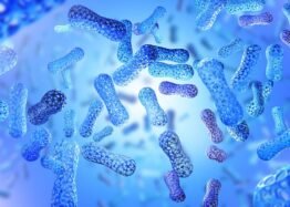
Therefore, the researchers decided to see how well probiotics could work as a therapeutic strategy. They used a human-origin probiotics cocktail consisting of five Lactobacillus and five Enterococcus strains that had previously been linked to reducing gut permeability and inflammation [4].

A cocktail for Alzheimer’s

The researchers used APP/PS1 mice, which are genetically modified to express human amyloid-β (Aβ). These mice develop signs of Alzheimer’s disease as the Aβ accumulates, and their cognitive abilities decrease earlier than those of wild-type mice.

In this experiment, 6- to 8-week-old APP/PS1 mice received a human-origin probiotic cocktail for 16 weeks. This treatment led to decreased Aβ accumulation in the hippocampal region of the brain, which is the first region where Alzheimer’s disease changes manifest, and mitigated the mice’s cognitive decline compared to untreated controls, suggesting that the treatment protected against the progression of Alzheimer’s disease.

Reduced inflammation

Apart from Aβ plaques, Alzheimer’s disease is also linked to neuroinflammation. Studies even suggest that systemic inflammation in mid-life can promote cognitive decline even 20 years later [5].

After giving their probiotic cocktail to mice, the researchers observed reduced neuroinflammation, decreased activation of the brain’s immune cells (microglia), and improved integrity of the blood-brain barrier, which regulates the entry of molecules and substances from blood to the brain. Systemic and gut inflammation were also reduced compared to controls, as measured by inflammatory markers in the blood and gut.

Better gut health

Probiotics seemed to have a broad positive impact on the gut that extended beyond inflammation. Testing of multiple markers of gut health showed improvements in the probiotic-treated animals compared to the control mice, such as reductions in gut permeability and structural and functional improvements to the linings of both the large and small intestines (intestinal epithelia). The researchers believe that the effectiveness of their probiotics cocktail is likely to be due to these improvements in intestinal barrier integrity.

As expected, the probiotic treatment had significant effects on the gut microbiomes of the treated mice. While this treatment didn’t impact microbial diversity, it affected the abundance of different microbial populations, increasing the numbers of beneficial microbes.

Males benefit more from probiotics

The risks of Alzheimer’s disease development and progression differ by sex. Therefore, the researchers examined differences between the data that they obtained from male and female mice.

They noted that while cognitive performance and reduction in Aβ were observed in both sexes, males had slightly better results than females. This was due to the fact that some, but not all, of the molecular mechanisms that provide such cognitive benefits differ between males and females.

The impact of probiotic treatment on microglial activation and inflammation was similar in male and female mice, except for one of the inflammatory markers in the brain (Il-1β), which was significantly reduced in male but not in female mice.

However, the researchers noted several positive changes in gut permeability, blood-brain barrier, and inflammation that were observed only in male mice but not females. They also noted that probiotic treatment had different impacts on the microbiomes of male and female mice.

Gut-brain connection

The researchers discussed possible mechanisms, looking at both previous research and their own results. They believe that an imbalance in gut microbes, specifically an increase in microbes associated with inflammation, leads to local gut inflammation that causes gut leakiness. This leads to a leakage of pro-inflammatory molecules into the blood, resulting in systemic inflammation that ultimately reaches the brain.

These pro-inflammatory molecules can harm the integrity of the blood-brain barrier, which allows them to infiltrate the brain and activate the brain immune system (microglia), leading to neuroinflammation. The researchers hold that this cascade from the gut to the brain contributes to the accumulation of Aβ and results in the progression of Alzheimer’s disease.

[Probiotic treatment] suppresses the origin of inflammation from the gut by preventing gut permeability, thereby keeping systemic inflammation in check. This, in turn, preserves the function of the BBB, preventing pro-inflammatory burdens on the brain and maintaining control over microglia activation, neuroinflammation, Aβ accumulation. Ultimately, this helps preserve cognitive health and protect against AD progression.

While this study shows a possible mechanism of the connection between the gut-brain axis and Alzheimer’s disease and provides supporting evidence, it still needs more data to prove that the proposed mechanism is correct. Further experiments and studies in different models of Alzheimer’s disease could enrich and support these conclusions, and safety and efficacy would need to be examined in human beings.

We would like to ask you a small favor. We are a non-profit foundation, and unlike some other organizations, we have no shareholders and no products to sell you. All our news and educational content is free for everyone to read, but it does mean that we rely on the help of people like you. Every contribution, no matter if it’s big or small, supports independent journalism and sustains our future.

Literature

[1] Prajapati, S. K., Wang, S., Mishra, S. P., Jain, S., & Yadav, H. (2025). Protection of Alzheimer’s disease progression by a human-origin probiotics cocktail. Scientific reports, 15(1), 1589.

[2] Vogt, N. M., Kerby, R. L., Dill-McFarland, K. A., Harding, S. J., Merluzzi, A. P., Johnson, S. C., Carlsson, C. M., Asthana, S., Zetterberg, H., Blennow, K., Bendlin, B. B., & Rey, F. E. (2017). Gut microbiome alterations in Alzheimer’s disease. Scientific reports, 7(1), 13537.

[3] Grabrucker, S., Marizzoni, M., Silajdžić, E., Lopizzo, N., Mombelli, E., Nicolas, S., Dohm-Hansen, S., Scassellati, C., Moretti, D. V., Rosa, M., Hoffmann, K., Cryan, J. F., O’Leary, O. F., English, J. A., Lavelle, A., O’Neill, C., Thuret, S., Cattaneo, A., & Nolan, Y. M. (2023). Microbiota from Alzheimer’s patients induce deficits in cognition and hippocampal neurogenesis. Brain : a journal of neurology, 146(12), 4916–4934.

[4] Ahmadi, S., Wang, S., Nagpal, R., Wang, B., Jain, S., Razazan, A., Mishra, S. P., Zhu, X., Wang, Z., Kavanagh, K., & Yadav, H. (2020). A human-origin probiotic cocktail ameliorates aging-related leaky gut and inflammation via modulating the microbiota/taurine/tight junction axis. JCI insight, 5(9), e132055.

[5] Walker, K. A., Gottesman, R. F., Wu, A., Knopman, D. S., Gross, A. L., Mosley, T. H., Jr, Selvin, E., & Windham, B. G. (2019). Systemic inflammation during midlife and cognitive change over 20 years: The ARIC Study. Neurology, 92(11), e1256–e1267.

Publié le Laisser un commentaire

Mitochondrial Damage May Drive Type 2 Diabetes

A new study suggests that damaged mitochondria activate the integrated stress response, which causes pancreatic β-cells, as well as liver and fat cells, to lose their identity and malfunction [1]. Blocking this response had benefits in mouse models.

The mitochondria-diabetes connection

Like with many diseases, the prevalence of type 2 diabetes grows with age. Therefore, age-related dysregulation of some kind contributes to the development of the disease. However, scientists have been struggling to unearth the exact causes.

The central feature of diabetes is the inability of β-cells that reside in the pancreas to produce insulin, which is needed to store glucose and maintain glucose homeostasis. To do their job, β-cells need energy, which comes from mitochondria. Mitochondrial dysfunction is a hallmark of aging, and since most cells have mitochondria, its impact on living organisms is wide and heterogeneous [2].

Mitochondrial dysfunction has long been linked to diabetes [3], but the causality direction remained unclear. Do failing mitochondria make beta cells worse at their job, or is it the other way around? In a new paper published in Science, researchers from the University of Michigan shed some light on this question, with potentially powerful implications for future therapies.

Cells from diabetic donors have bad mitochondria

First, the scientists confirmed that human clusters of pancreatic endocrine cells (islets), including β-cells, from type 2 diabetes patients bear signs of mitochondrial dysfunction. Beta cells, but no other types of cells, from these patients had less mitochondrial DNA (mtDNA) and lower expression of 11 of 13 mitochondrial resident genes than in healthy controls. Mitophagy, the process of discarding malfunctioning mitochondria, was impaired as well.

These findings pointed at serious problems with the mechanisms of mitochondrial quality control. Interestingly, cells from simply obese donors or donors with insulin resistance did not show the same level of mitochondrial dysfunction, suggesting that this mitochondrial quality control loss was specific to diabetes patients.

How immature of you, β-cells

To see if impairing mitochondrial quality control can induce β-cell failure, the researchers engineered three mouse models with different mitochondrial pathways rendered deficient. The first model featured deletion of CLEC16A, a regulator of mitophagy, the second had reduced mtDNA content due to loss of TFAM, a regulator of mitochondrial genome integrity, and in the third, Mitofusins 1 and 2, proteins that promote mitochondrial fusion, were knocked out.

In all three models, messing with mitochondria triggered the integrated stress response (ISR). ISR is a cellular signaling network that is activated across various cell types to manage stress and maintain homeostasis by tweaking protein production. However, when persistently engaged, it can negatively impact cellular function. As they dug deeper to discover exactly how, the researchers received a surprise.

Apparently, mitochondria-to-nucleus (retrograde) ISR signaling dampened the expression of transcription factors that are central for β-cell maturity, identity, and function. As a result, the affected β-cells became less differentiated than their healthy counterparts.

“We wanted to determine which pathways are important for maintaining proper mitochondrial function,” said Dr. Emily M. Walker, a research assistant professor of internal medicine and first author of the study. “In all three cases, the exact same stress response was turned on, which caused β-cells to become immature, stop making enough insulin, and essentially stop being β-cells.”

Jamming the signal brings the cells back

Diabetes affects other metabolic tissues, such as liver tissue, muscle, and fat. To investigate further, the researchers ran similar experiments in mouse models of impaired mitochondrial quality control in liver cells (hepatocytes) and brown fat cells (adipocytes), with similar results.

“Diabetes is a multi-system disease: you gain weight, your liver produces too much sugar, and your muscles are affected. That’s why we wanted to look at other tissues as well,” said Scott A. Soleimanpour, M.D., director of the Michigan Diabetes Research Center and senior author of the study. “Although we haven’t tested all possible cell types, we believe that our results could be applicable to all the different tissues that are affected by diabetes.”

Can this be fixed? Several years ago, a potent ISR blocker called ISRIB was discovered and is currently in several clinical trials, including by Alphabet’s company Calico and the pharma giant AbbVie. The researchers treated mouse islets with ISRIB and found that it robustly restores β-cell identity markers specifically by inhibiting retrograde ISR signaling.

“Losing your β-cells is the most direct path to getting type 2 diabetes. Through our study we now have an explanation for what might be happening and how we can intervene and fix the root cause,” Soleimanpour said.

A possible basis for new treatments

Some other mitochondria researchers founded the study intriguing. “Using diet and genetic manipulations, the authors show the importance of robust mitophagy and retrograde signaling from mitochondria to nucleus in the involvement of mitochondrial function in type 2 diabetes,” said Dr. Amutha Boominathan, head of mitochondria research at the Longevity Research Institute. Boominathan and her team recently published an exciting study on nuclear expression of mitochondrial genes.

“What is interesting,” she added, “is that the authors find converging pathways in the β-Clec16aKO, β-Mfn1/2DKO and the Tfam-deficient mice in triggering the mitochondrial ISR influencing tissue specific blockade in cell differentiation not only in pancreas but also in other metabolic tissues such as liver and adipose tissues. The authors systematically address the causality for the role mitochondria play in age-associated metabolic diseases such as type 2 diabetes.”

Dr. Spring Behrouz, CEO of Vincere Biosciences, a mitochondria-targeting longevity biotech company, was impressed by the new study as well. “Reduced mtDNA levels, disrupted mitochondrial structure, and impaired mitophagy in metabolic tissues are often seen as secondary effects of other factors in type 2 diabetes,” she said. “However, this data suggests that early mitochondrial dysfunction actively contributes to the disease and potentially impacts other tissues.”

According to Behrouz, the study’s findings might be important for developing new mitochondria-based therapies. “By demonstrating the impact of mitochondrial quality control in metabolic tissue identity, this research opens up entirely new possibilities for treatment of diabetes as well as a range of other metabolic disorders,” she said.

We would like to ask you a small favor. We are a non-profit foundation, and unlike some other organizations, we have no shareholders and no products to sell you. All our news and educational content is free for everyone to read, but it does mean that we rely on the help of people like you. Every contribution, no matter if it’s big or small, supports independent journalism and sustains our future.

Literature

[1] Walker, E. M., Pearson, G. L., Lawlor, N., Stendahl, A. M., Lietzke, A., Sidarala, V., … & Soleimanpour, S. A. (2025). Retrograde mitochondrial signaling governs the identity and maturity of metabolic tissues. Science, eadf2034.

[2] Srivastava, S. (2017). The mitochondrial basis of aging and age-related disorders. Genes, 8(12), 398.

[3] Kwak, S. H., Park, K. S., Lee, K. U., & Lee, H. K. (2010). Mitochondrial metabolism and diabetes. Journal of diabetes investigation, 1(5), 161-169.

Publié le Laisser un commentaire

Creating a Functional Pancreas From Human Cells

In Cell Reports Medicine, researchers have described how they created a fully functional pancreas made from human cells that has been found to work in mice.

A new era of organ replacement

In their introduction, the researchers discuss the well-known problems with insulin injections to treat Type 1 diabetes: the sort of constant monitoring that is required is difficult for patients to consistently comply with [1], and daily manual injections can’t adequately simulate the responsiveness of pancreatic tissue [2]. Direct injection of beta islet cells, which produce insulin, are limited by donor organs and require the immune system to be suppressed [3].

More modern techniques recognize that the extracellular matrix (ECM) governs a large part of how stem cells differentiate [4], and the effects of the ECM on the pancreas have been investigated in detail [5]. This led to a rapid increase in the work being done in this area, with ECM structures being created for the purpose [6]. The researchers of this paper had created a functional pancreas with insulin-producing cells from pigs, and it was functional in mice [7].

Therefore, the next step was to use cells derived from human beings.

Constructed organs are more effective than previous approaches

The researchers used two separate kinds of cells derived from human induced pluripotent stem cells (iPSCs): insulin-producing islet cells (SC-islets) and endothelial cells (iECs), which line the walls of arteries and veins. First, the researchers brought together these cells in a 9-to-1 ratio in order to produce spheroids (ViβeSs). Then, they populated decellularized rat lung tissue with more iECs and let them grow for two days, and finally, they injected this ready tissue with ViβeSs and more iECs to promote blood vessel formation (vascularization), creating a vascularized endothelia pancreas built from iPSCs: an iVEP.


This approach worked. The injected ViβeSs did not come loose from the structure; instead, the iECs formed vascular tissue within the decellularized lungs, providing them with stability and the blood flow that they need to perform their duties. The iVEP structure was found to grant significant improvements to the cells’ survival and responsiveness, with the cells producing more insulin under high-glucose conditions.

In immunocompromised diabetic mice, the iVEP structure also performed much better than ViβeSs put into a a pre-vascularized pouch under the skin. In only two out of the thirteen mice given the latter, normal glycemia was established within a month; this happened in all the mice given iVEPs. Half of the iVEP-receiving mice had normal glycemia within two weeks of transplantation.

Unsurprisingly, removing the iVEPs from these mice led to diabetes within a week. An exaination of these structures revealed extensive vascular connections: the mice had successfully integrated the iVEPs into their bodies. Further investigation found that iECs were necessary in the creation of iVEPs; without endothelial cells, the structures fail to properly integrate into the vascular structure.

Decellularized, vascularized structures as a way forward

With these results in hand, the researchers compared their iVEP approach to previous work. They hold that their approach to vascularization improves many fundamental aspects of cellular development: for example, they note that by themselves, islet cells generated from iPSCs require 20 days to reach a mature and effective phenotype [8]. Meanwhile, in iVEPs, the cells require only a week to reach this phenotype.

While the researchers note that their approach is more complicated than products that are already in clinical trials, they believe that it will make for a better product. However, their existing scaffolds, derived from rats, are far too small to use in people. They plan to use pig organs instead, and they hope to use hypo-allergenic cells that completely get rid of the risk of immune rejection and the need for potentially risky immunosuppressants.

The researchers developed this approach to treat type 1 diabetes, but this work has implications for many other diseases, many of which are age-related. While replacing the pancreas cannot heal the insulin resistance inherent in type 2 diabetes, it may be a viable strategy for long-term loss of function. Similarly, this technology can potentially be applied to many other organs, including the lungs and heart. While wholesale replacement of human organs with bioengineered equivalents is still not on the table, this technology continues to advance to the clinic.

We would like to ask you a small favor. We are a non-profit foundation, and unlike some other organizations, we have no shareholders and no products to sell you. All our news and educational content is free for everyone to read, but it does mean that we rely on the help of people like you. Every contribution, no matter if it’s big or small, supports independent journalism and sustains our future.

Literature

[1] Beck, R. W., Bergenstal, R. M., Laffel, L. M., & Pickup, J. C. (2019). Advances in technology for management of type 1 diabetes. The Lancet, 394(10205), 1265-1273.

[2] Piemonti, L. (2021). Felix dies natalis, insulin… ceterum autem censeo “beta is better”. Acta Diabetologica, 58(10), 1287-1306.

[3] Pepper, A. R., Bruni, A., & Shapiro, A. J. (2018). Clinical islet transplantation: is the future finally now?. Current opinion in organ transplantation, 23(4), 428-439.

[4] Hogrebe, N. J., Augsornworawat, P., Maxwell, K. G., Velazco-Cruz, L., & Millman, J. R. (2020). Targeting the cytoskeleton to direct pancreatic differentiation of human pluripotent stem cells. Nature biotechnology, 38(4), 460-470.

[5] Berger, C., Bjørlykke, Y., Hahn, L., Mühlemann, M., Kress, S., Walles, H., … & Zdzieblo, D. (2020). Matrix decoded–A pancreatic extracellular matrix with organ specific cues guiding human iPSC differentiation. Biomaterials, 244, 119766.

[6] Peloso, A., Urbani, L., Cravedi, P., Katari, R., Maghsoudlou, P., Fallas, M. E. A., … & Orlando, G. (2016). The human pancreas as a source of protolerogenic extracellular matrix scaffold for a new-generation bioartificial endocrine pancreas. Annals of surgery, 264(1), 169-179.

[7] Citro, A., Neroni, A., Pignatelli, C., Campo, F., Policardi, M., Monieri, M., … & Piemonti, L. (2023). Directed self-assembly of a xenogeneic vascularized endocrine pancreas for type 1 diabetes. Nature Communications, 14(1), 878.

[8] Velazco-Cruz, L., Song, J., Maxwell, K. G., Goedegebuure, M. M., Augsornworawat, P., Hogrebe, N. J., & Millman, J. R. (2019). Acquisition of dynamic function in human stem cell-derived β cells. Stem cell reports, 12(2), 351-365.